Radiological identification and analysis of soft tissue musculoskeletal calcifications, Insights into Imaging
Por um escritor misterioso
Last updated 07 setembro 2024
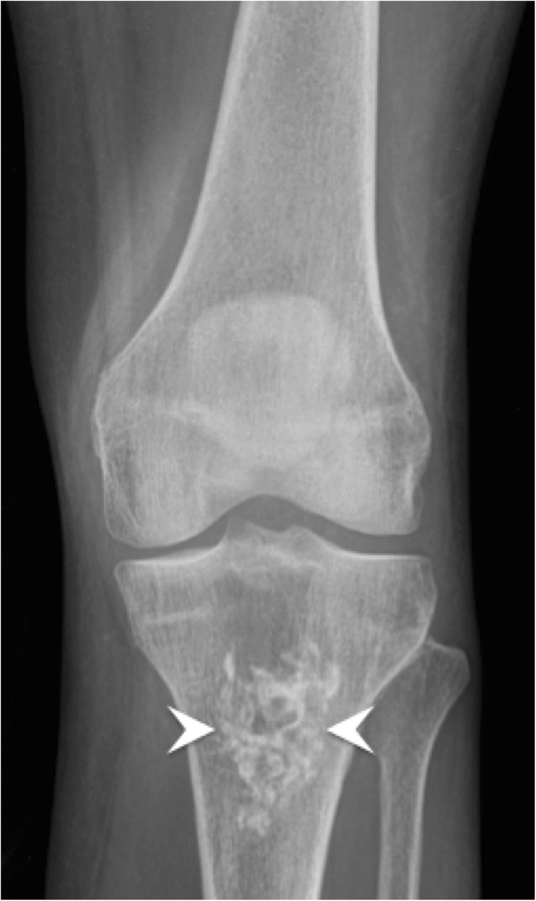
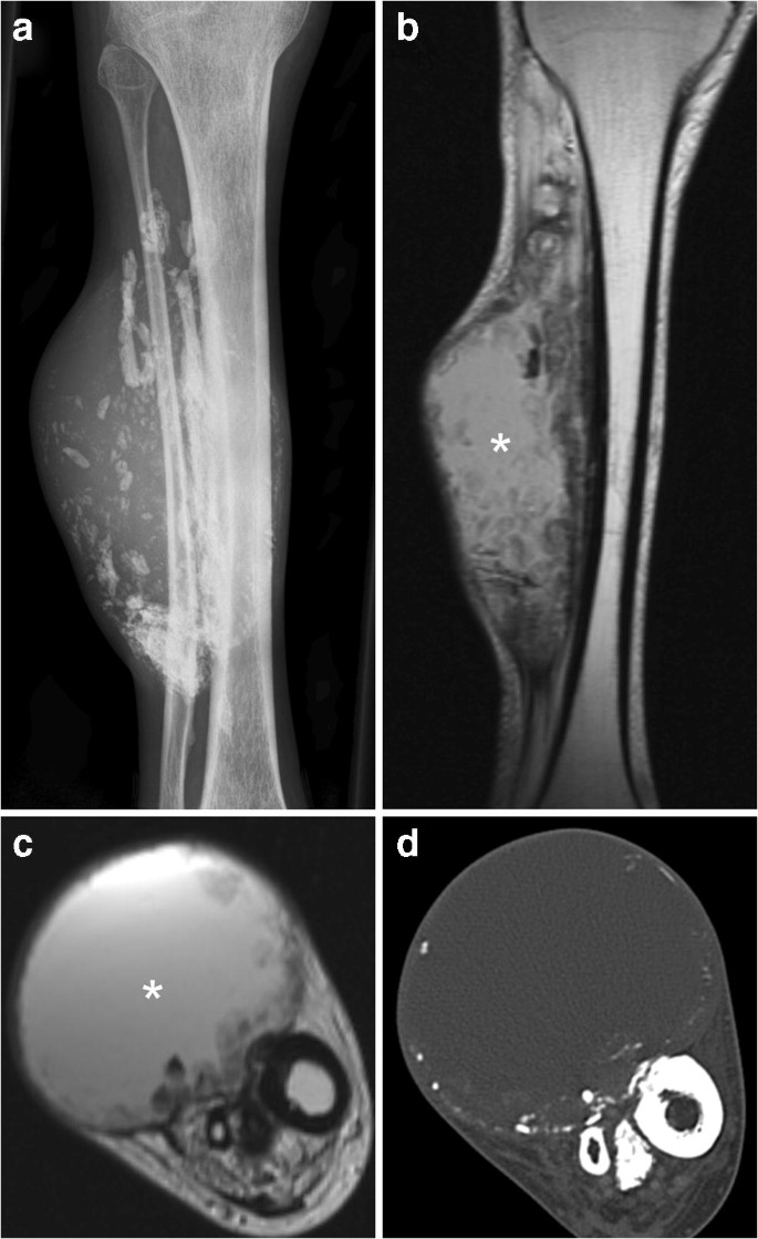
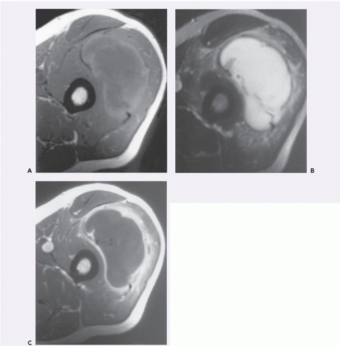
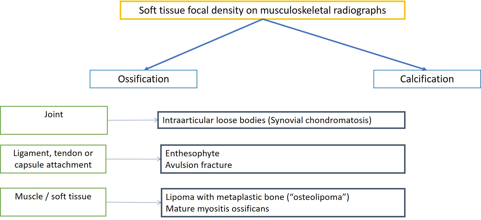
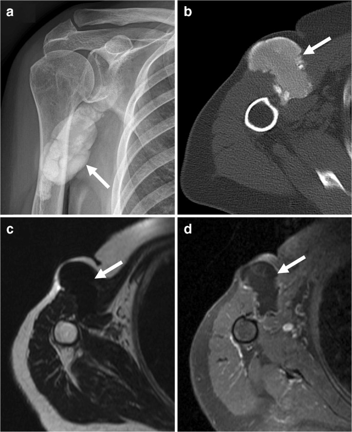
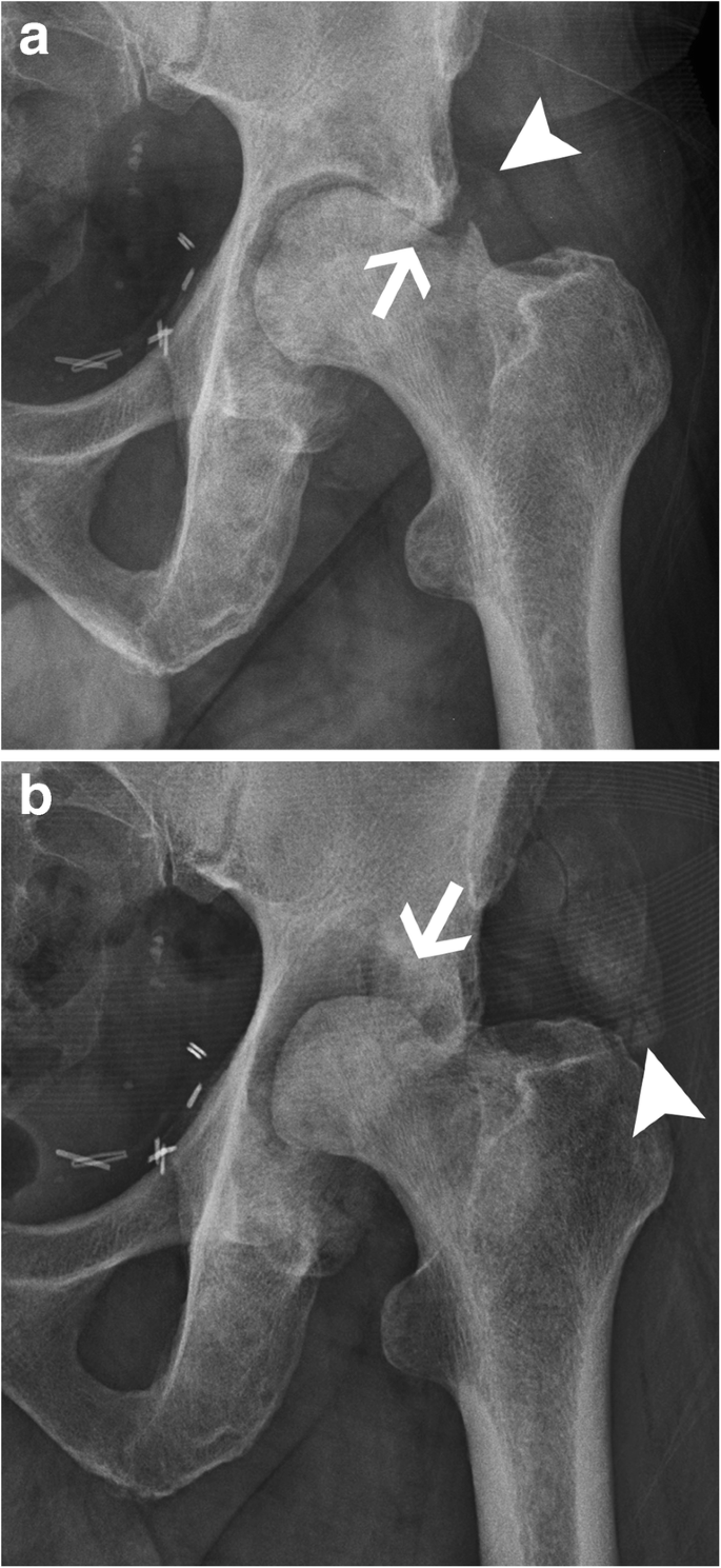
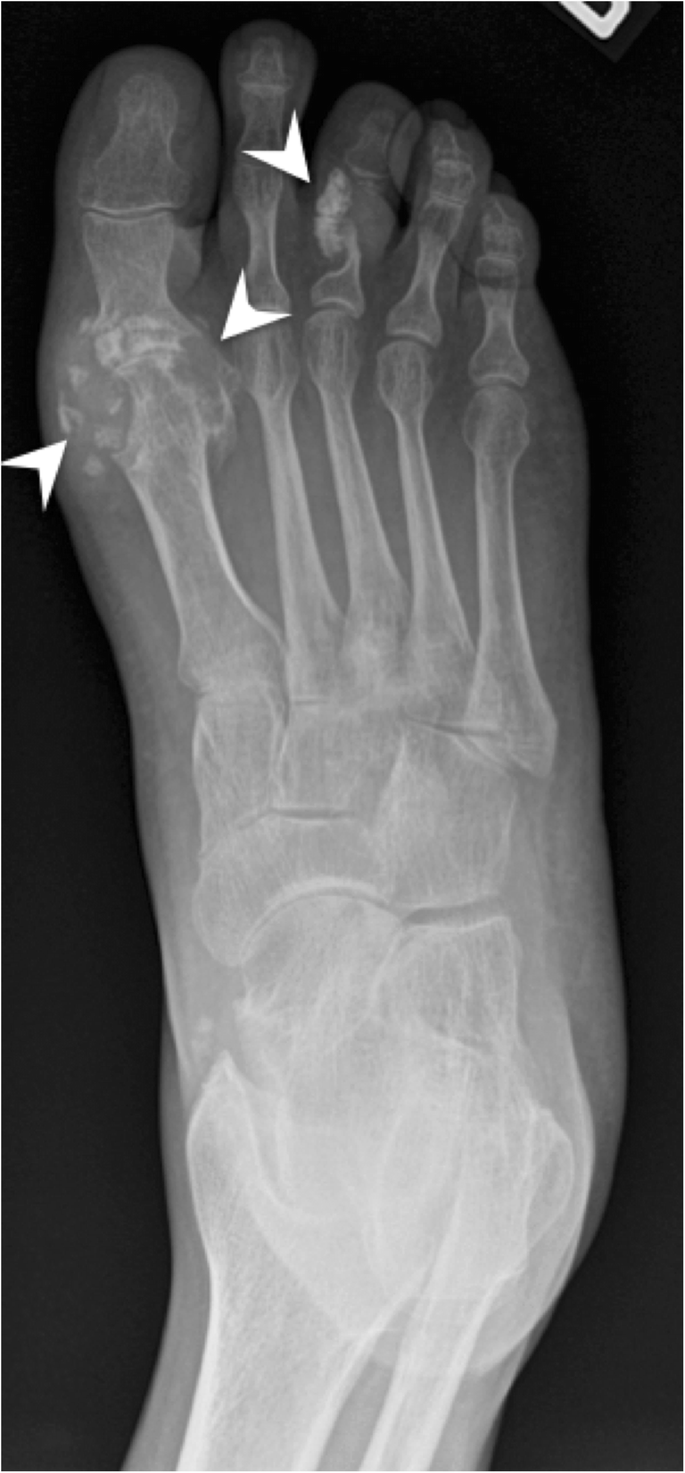
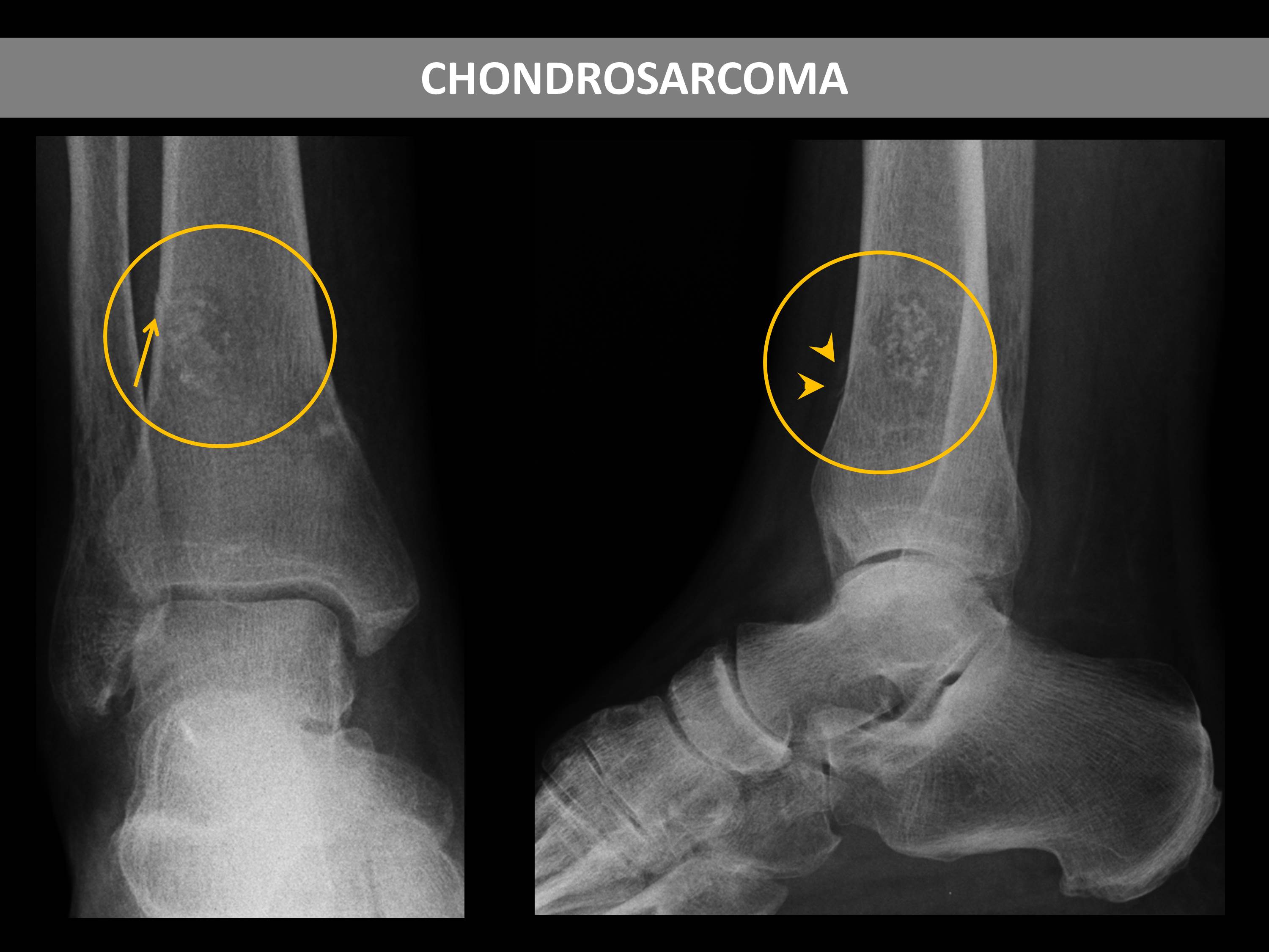
Abstract Musculoskeletal calcifications are frequent on radiographs and sometimes problematic. The goal of this article is to help radiologists to make the correct diagnosis when faced with an extraosseous musculoskeletal calcification. One should first differentiate a calcification from an ossification or a foreign body and then locate the calcification correctly. Each location has a specific short differential diagnosis, with minimal further investigation necessary. Intra-tendon calcifications are most frequently associated with hydroxyapatite deposition disease (HADD). In most cases, intra-articular calcifications are caused by calcium pyrophosphate dihydrate (CPPD) crystal deposition disease. Soft tissue calcification can be caused by secondary tumoural calcinosis from renal insufficiency, or collagen vascular diseases and by vascular calcifications, either arterial or venous (phlebolith). Teaching Points • Calcifications have to be differentiated form ossification and foreign body. • A musculoskeletal MRI study must always be correlated with a radiograph. • The clinical manifestations of calcifications may sometimes mimic septic arthritis or sarcoma. • HADD and CPPD crystal deposition have a distinct appearance on radiograph. • Calcinosis is more frequently caused by chronic renal failure and scleroderma.
Musculoskeletal Archives - UCSD Ultrasound

Ultrasound Appearance of the Migration of Tendon Calcifications - Bianchi - 2019 - Journal of Ultrasound in Medicine - Wiley Online Library
Calcified or ossified benign soft tissue lesions that may simulate malignancy
Imaging of Soft Tissue Masses

Microorganisms, Free Full-Text
EPOS™ - C-07775

SciELO - Brasil - Soft tissue calcifications: a pictorial essay Soft tissue calcifications: a pictorial essay
Calcified or ossified benign soft tissue lesions that may simulate malignancy

Radiological identification and analysis of soft tissue musculoskeletal calcifications. - Abstract - Europe PMC
Radiological identification and analysis of soft tissue musculoskeletal calcifications, Insights into Imaging

Hydroxyapatite Deposition Disease (HADD) of the Greater Trochanter
Radiological identification and analysis of soft tissue musculoskeletal calcifications, Insights into Imaging
Recomendado para você
-
 In the tibia hi-res stock photography and images - Alamy07 setembro 2024
 -
 Tibialis Band Portable Tibialis Trainer Calf Raise Machine, Upgrade Tibia Dorsi Calf Machine with Resistance Bands, Professional Tibiali Anterior Exercise Equipment for Leg Calves Shins Strengthener : Sports & Outdoors07 setembro 2024
 -
 Is there some trick to managing plasma rings? : r/TibiaMMO07 setembro 2024
 -
 Outcast Open-Tibia 7.6 - The Ultimate Wiki — Outcast Wiki Latest documentation07 setembro 2024
 -
 Figure 3 from Correction of genu recurvatum by the Ilizarov method.07 setembro 2024
 -
EPOS™07 setembro 2024
-
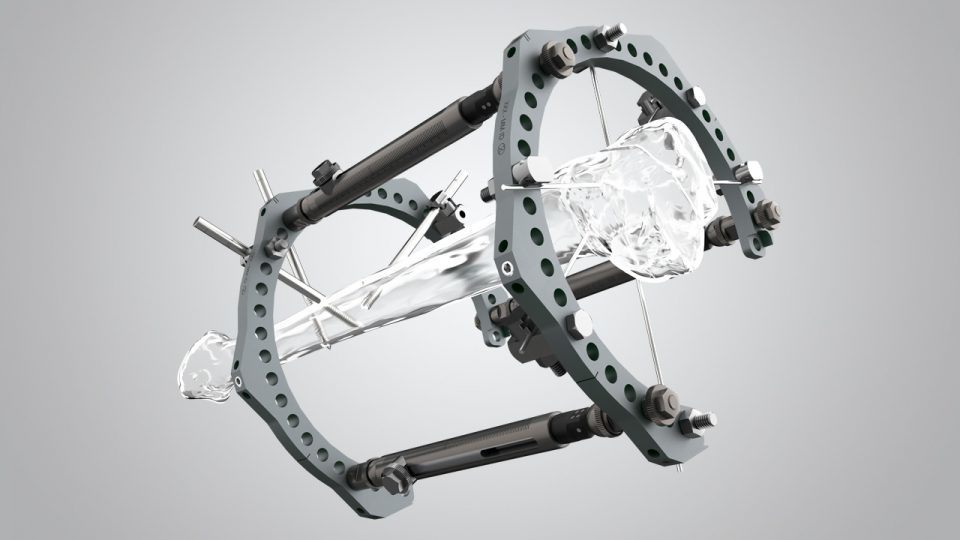 TL-HEX TrueLok Hexapod System - Orthofix07 setembro 2024
 -
 Tibia's Summons Elden Ring Wiki07 setembro 2024
 -
 McKesson Brand 155-81-82397 - McKesson Medical-Surgical07 setembro 2024
 -
 Medical Ring Fixator for Tibial & Femur Fracture in Orthopedic Tibia Industry - China Ilizarov, Ortopedi Ilizarov07 setembro 2024
você pode gostar
-
 GG large tote bag07 setembro 2024
 -
 Send Engage Surveys to Specific Audiences : Limeade Listening07 setembro 2024
 -
.jpg) Gaelic Games - Football (Europe) ROM (ISO) Download for Sony Playstation 2 / PS207 setembro 2024
Gaelic Games - Football (Europe) ROM (ISO) Download for Sony Playstation 2 / PS207 setembro 2024 -
 Selects Mario Party: Island Tour 3DS - Compra jogos online na07 setembro 2024
 -
 Mu Matrix Season 6.3 - Publica tu servidor - Creando Mu Online07 setembro 2024
 -
 Casa Móbile Mega Store - Móveis e Decoração07 setembro 2024
 -
Page 295 - ALL Roblox Clothing Codes (Pants, Shirts, T-Shirts07 setembro 2024
-
 Assistir Saint Seiya - Soul of Gold (Os Cavaleiros do Zodíaco07 setembro 2024
 -
 Sonic the Hedgehog 3 - Chaos Emeralds Chaos emeralds, Sonic the hedgehog, Sonic07 setembro 2024
 -
 20 Best iPhone, iPad, Android and Windows Phone games this week, Games07 setembro 2024