Echo image of right ventricular apex (RVA) pacemaker, pressure
Por um escritor misterioso
Last updated 10 novembro 2024


Computed tomography validated right ventricular mid‐septal lead implantation using right ventricular angiography - Shenthar - 2021 - Journal of Arrhythmia - Wiley Online Library

Echo image of right ventricular apex (RVA) pacemaker, pressure

Advantage of right ventricular outflow tract pacing on cardiac function and coronary circulation in comparison with right ventricular apex pacing.

Subacute right ventricular pacemaker lead perforation: evaluation by echocardiography and cardiac CT
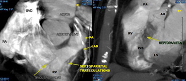
A proposed technique for right ventricular septal pacing

An X-ray example of typical positioning of RVOT lead in anteroposterior

Final position of the wire positioned in the apex of the right
Acute Impact of Pacing at Different Cardiac Sites on Left Ventricular Rotation and Twist in Dogs
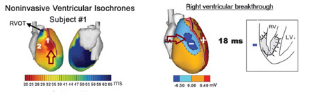
A proposed technique for right ventricular septal pacing

Figure. Exemplificative diagram for RVOT septal and RVAP pacing. Chest

Echo image of right ventricular apex (RVA) pacemaker, pressure

PDF) Hisian area and right ventricular apical pacing differently affect left atrial function: An intra-patients evaluation
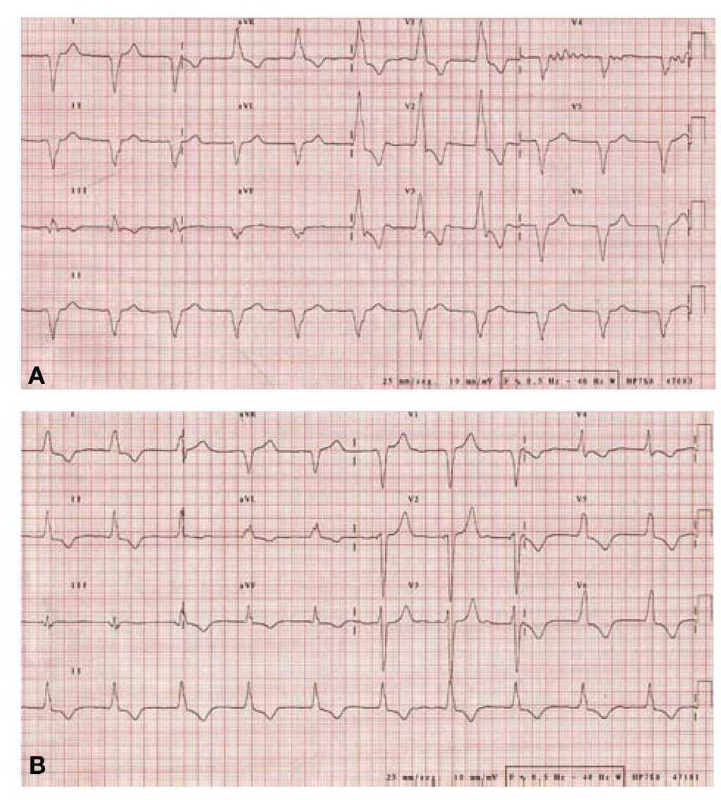
Left Ventricular Pacing. Is It Always Better Than Right Ventricular Pacing?
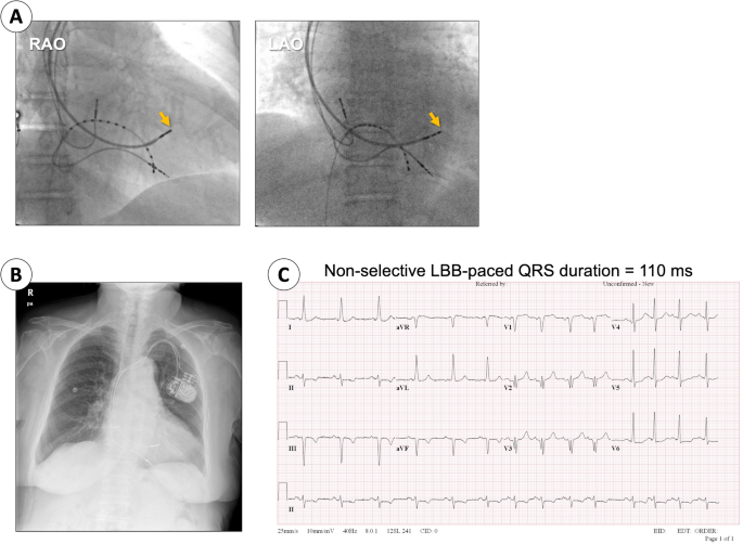
Reversal of pacing-induced cardiomyopathy after left bundle branch area pacing: a case report, International Journal of Arrhythmia
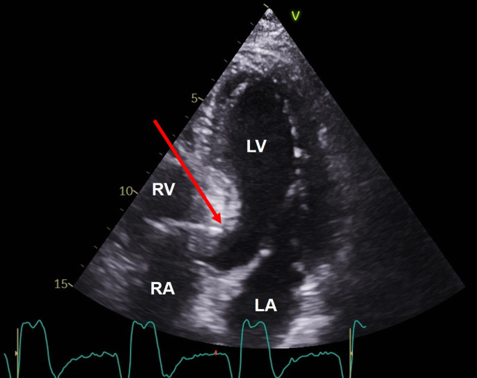
Anatomy for right ventricular lead implantation Herzschrittmachertherapie + Elektrophysiologie
Recomendado para você
-
 Ezequiel 33:11 RVA - Diles: Vivo yo, dice el Señor Jehová, que no10 novembro 2024
 -
The Sanctuary Colonial Heights VA10 novembro 2024
-
 Baile Reviver com 'Os Mineirinhos' - Fundação Cultural de Casimiro10 novembro 2024
 -
 Qué Pasa? Festival is Rescheduled Due to Weather - RVAHub10 novembro 2024
 -
 In Vivo University10 novembro 2024
 -
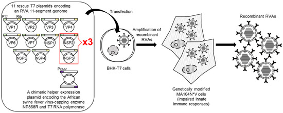 Viruses, Free Full-Text10 novembro 2024
 -
 UNI-VERSO: Entrevista na RVA - Rádio Venâncio Aires10 novembro 2024
 -
 RVA Latinofest – Sacred Heart Parish10 novembro 2024
 -
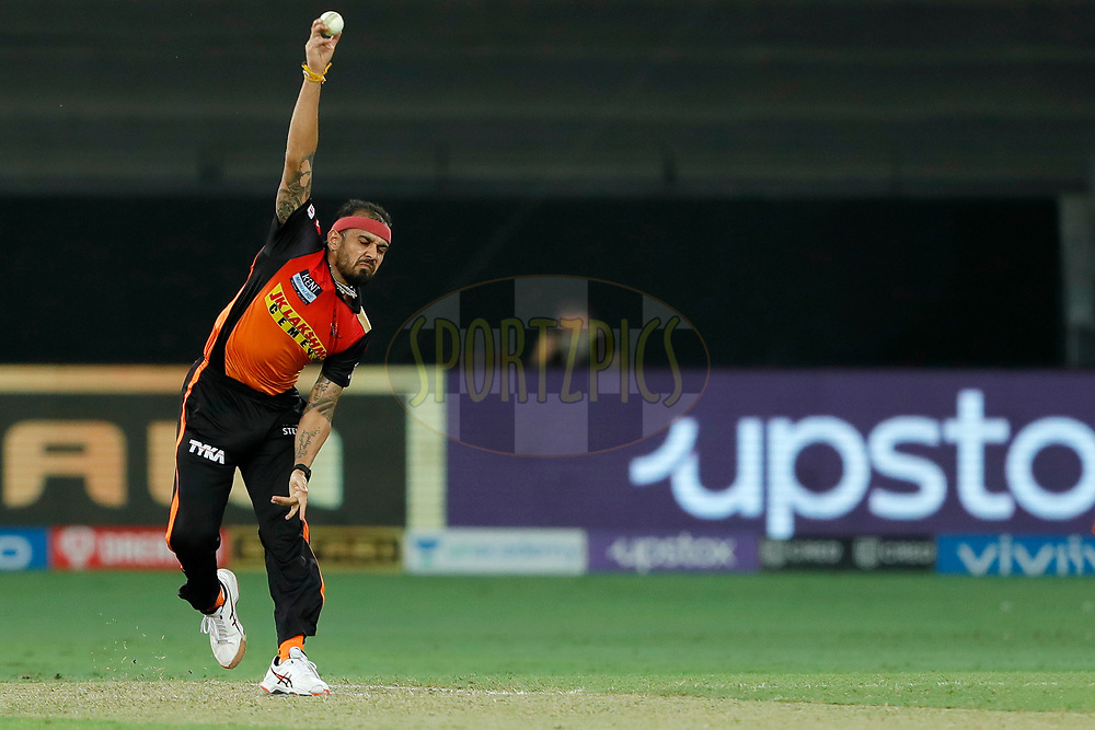 Vivo IPL M49 - KKR v SRH10 novembro 2024
 -
 Viva RVA! — Creative Agency in Richmond VA, One Man Design10 novembro 2024
você pode gostar
-
Holder Chithambaram and Vahap share Masters division lead at Dubai chess meet - GulfToday10 novembro 2024
-
 Jogador De Futebol Que Corre O Fundo Da Bola. Imagem Baixa Do Ângulo Da Bola De Pontapé Do Menino Do Futebol No Campo De Treinamento Da Grama Foto Royalty Free, Gravuras, Imagens10 novembro 2024
 -
 ❄️🍂🌿🐱-Dark Piece-🐱🌿🍂❄️ on X: Cute little Horror from 2019 >:3 #UndertaleAU #Sans #Horrortale #Horror_Sans #HorrorSans #Art / X10 novembro 2024
 -
 Hitomi Flor - Descendientes 3 - Rotten to the Core (D3 Remix) (Cover en Español): listen with lyrics10 novembro 2024
 -
 Fogos de artifício, jogos pirotécnicos para celebrar o ano novo ou10 novembro 2024
 -
 Ranking the Sun God Luffy's Gears10 novembro 2024
 -
PSN Gift Cards Codes Contest - Apps on Google Play10 novembro 2024
-
 CQNET Friends Baby Monthly Fleece Milestone Growth Blanket, Could I be Any Cuter? Friends Frame Newborn Baby Milestone Blanket, Pivot Age Blanket Baby Present Friends Theme TV Show Merchandise Large: Buy Online10 novembro 2024
 -
 What's the best game that's uncomfortably pro-monarchy? The Best10 novembro 2024
 -
 AO VIVO: Assista a Flamengo x Bragantino com o Coluna do Fla10 novembro 2024

